What is ureteric stenting?
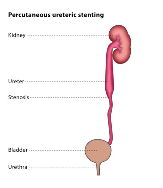
Ureteric stents enter the body through a tiny hole in your back. They are used when urine from the kidney is blocked and cannot pass normally through the ureter into the bladder. This condition might occur because of a blockage caused by a stone, scarring, infection, or unusual tissue growth. When urine cannot flow normally, it can lead to discomfort, infections, and potentially harm the kidneys.
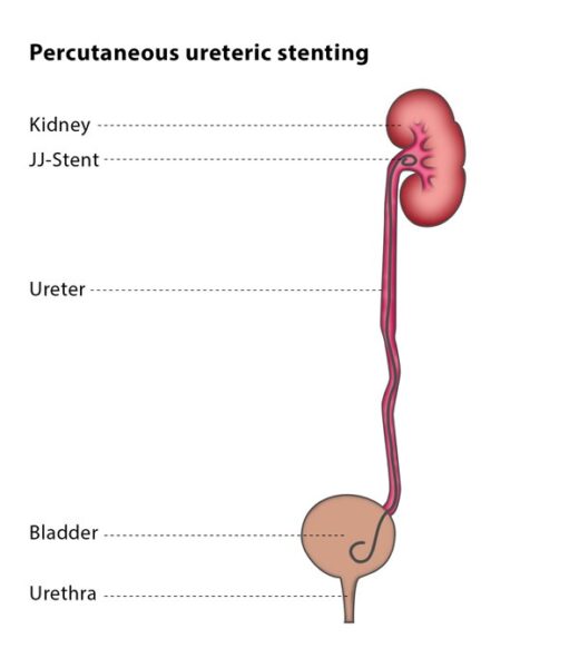
A ureteric stent (also called a J-J stent or double-J stent) is a thin, flexible plastic tube which is curled at both ends to avoid damaging the kidney and urinary bladder and to prevent it from dislocating. The stent is placed so that its upper end is in the kidney and its lower end is in the urinary bladder.
In some cases, these tubes are a temporary solution while the underlying issue causing the blockage of urine is addressed. This period might last several weeks or months. However, for others, these tubes are a more long-term necessity, requiring the stents to be replaced every few months.